Human Physiology Equipment
Clear demonstration tools make a major difference when teaching how the human body works. In classrooms, training labs, and introductory health science programs, learners often understand physiological concepts faster when they can connect structure, function, and observable effects through hands-on models and teaching aids. This is where Human Physiology Equipment becomes especially useful.
This category brings together educational equipment designed to support lessons on organs, body systems, sensory function, anatomy, and health awareness. It is well suited to schools, universities, training centers, and laboratory education environments that need practical resources for explaining complex biological processes in a more visual and memorable way.
Supporting physiology education through visual and practical learning
Human physiology is easier to teach when students can see relationships between body structures and their functions. Instead of relying only on diagrams or text, instructors can use anatomical models, charts, and simulation tools to explain topics such as the nervous system, sensory organs, reproduction, internal organs, and the effects of impaired perception.
In many teaching environments, these materials are used to reinforce core lessons, support lab-based exercises, and encourage discussion. For broader program needs, institutions may also combine this category with cell biology equipment when moving from microscopic structure to whole-body function.
Typical equipment found in this category
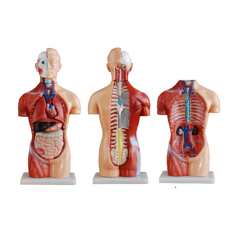
The range of products in this category is centered on educational physiology models and demonstration aids. These include organ models, body section models, anatomical charts, and teaching tools that help students identify structures and understand how different systems are organized.
Examples from PHYWE include the MOD-BRAIN Brain Model, MOD-EYE Eye Model, MOD-SKIN Skin Model, and the MOD-MINITORSO Mini Torso. These products are useful for demonstrating internal organization, orientation of organs, and the relationship between anatomical parts in a way that is more interactive than static illustrations alone.
There are also topic-specific models such as the MOD-MALEPELVIS Male Pelvis and MOD-FEMALEPELVIS Human Female Pelvis Section, which support teaching in reproductive anatomy and pelvic structure. For classroom wall reference, the ERL-AL163 Chart Internal Organs can complement physical models by giving students a large visual overview during lectures and guided explanations.
Health awareness and sensory impairment simulation
Not every physiology lesson is limited to anatomy. Some training programs also need tools that help learners understand how body function and perception can be affected by harmful substances. In this context, simulation glasses provide a practical way to demonstrate altered balance, spatial awareness, visual distortion, and reduced concentration.
Products such as the PHYWE KLA-110-205 Alcohol Intoxication Simulation Glasses, the KLA-110-207 version for stronger intoxication effects, and the KLA-110-209 Drugs Intoxication Simulation Glasses are relevant for safety education, health awareness sessions, and school-based prevention programs. Rather than presenting intoxication as an abstract concept, these tools help demonstrate how impaired perception can influence coordination and judgment.
This type of equipment can be particularly useful in interdisciplinary teaching, where physiology overlaps with health education, behavioral awareness, and public safety. Instructors can build realistic exercises around movement, visual tasks, and reaction challenges to make the lesson more engaging and easier to remember.
How to choose the right human physiology teaching equipment
The right selection depends largely on the level of instruction and the learning objective. If the goal is to teach basic organ identification and body organization, compact torso models, organ charts, and removable-part anatomical models are often the most efficient starting point. If the lesson focuses on sensory systems, dedicated models of the eye, brain, skin, or dental structures may be more appropriate.
For practical school use, it is helpful to consider how often the equipment will be handled, whether it needs to be moved between classrooms, and how much detail students actually need. A highly detailed model may be ideal for advanced teaching, while a more accessible format can be better for middle school or introductory courses. Programs that also teach system-level body functions may benefit from exploring related ecology and environment equipment when connecting human biology to broader life science topics.
Applications in schools, labs, and training centers
This category is relevant across a wide range of educational settings. Secondary schools often use these tools to support biology classes, especially when introducing body systems, sense organs, hygiene, and health education. Colleges and vocational institutions may use them for more focused training in healthcare, laboratory science, or science teaching methodology.
Models such as the MOD-TEETH Dental Care Model can support oral hygiene instruction, while the Brain Model and Eye Model are suitable for demonstrating specialized organs and their functions. The Student Set General Biology for grades 7-10 can also serve as a complementary resource for broader life science activities where physiology is taught alongside plants, nutrition, senses, and basic experimental work.
When course content extends beyond human systems into organism classification or comparative biology, users may also review zoology equipment or botany equipment to build a more complete biology teaching setup.
Why anatomical models remain important in physiology teaching
Even with digital learning resources widely available, physical teaching models still offer strong educational value. They allow students to observe depth, orientation, relative size, and removable parts in a way that is often more intuitive than a flat screen or textbook image. This is particularly important when introducing spatial anatomy and system relationships.
Physical interaction also supports group discussion and demonstration-based teaching. An instructor can point out structures directly, compare parts side by side, and guide students through functional explanations step by step. In practice, this makes physiology teaching more concrete and improves retention for many learners.
Building a more effective physiology learning environment
A well-planned teaching setup usually combines several formats rather than relying on one type of equipment alone. Anatomical charts provide overview, removable models show internal arrangement, and simulation tools create applied learning scenarios. Together, these resources help connect visual recognition with functional understanding.
Whether the requirement is a compact model for routine classroom use or a broader selection for a training laboratory, this category supports structured and practical biology education. Choosing the right mix of models, charts, and simulation aids can make lessons on human structure and function easier to teach, easier to understand, and more relevant to real-world learning outcomes.
-
-
-
-
-
-
-
-
-
-
-
-
-
-
-
-
-
-
-
-
-
-
-
-